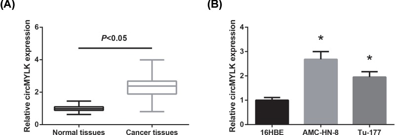
Figure 1. circMYLK is overexpressed in LSCC.
(A) circMYLK expression levels were validated in LSCC tissues and matched adjacent non-tumorous tissues by RT-qPCR analysis. (B) Expression levels of circMYLK in human LSCC cell lines and normal 16HBE cells were analyzed by RT-qPCR analysis. *P<0.05 vs. 16HBE cells.

