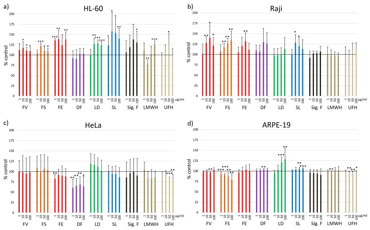
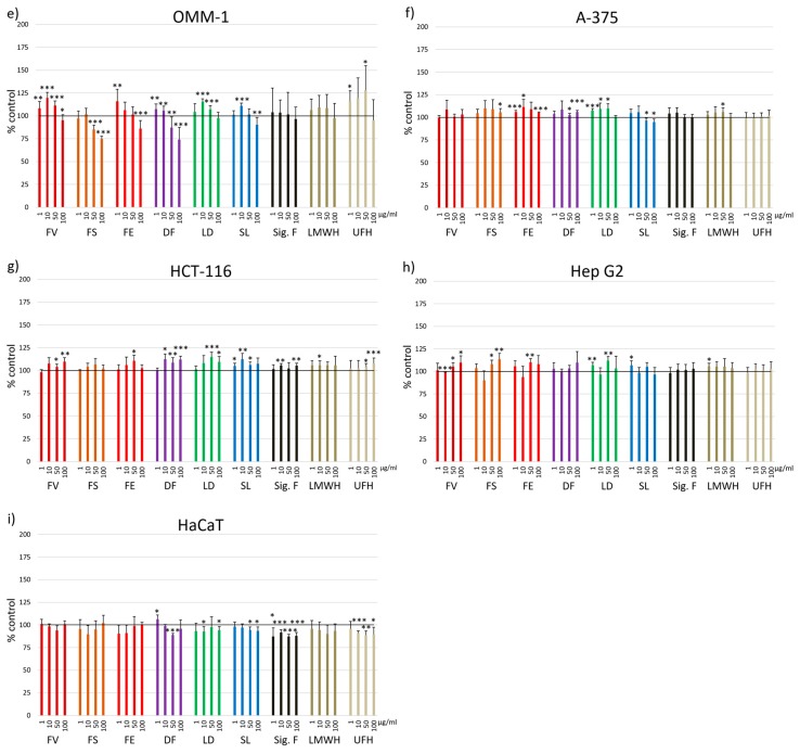
Figure 1.
Cell viability was determined using MTS assay after 24 h incubation with 1, 10, 50 and 100 µg/mL fucoidan from Fucus vesiculosus (FV), Fucus serratus (FS), Fucus distichus subsp. evanescens (FE), Dictyosiphon foeniculaceus (DF), Laminaria digitata (LD) and Saccharina latissima (SL) as well as the reference substances Sigma-Aldrich fucoidan (F. vesiculosus) (Sig. F), enoxaparin (LMWH) and heparin (UFH). Nine different cell lines were tested: a) HL-60, b) Raji, c) HeLa, d) ARPE-19, e) OMM-1, f) A-375, g) HCT-116, h) Hep G2 and i) HaCaT. Values are expressed as mean and standard deviation in relation to untreated cells (100%). Significances compared to control were determined using student’s t-test * p < 0.05, ** p < 0.01, *** p < 0.001, n ≥ 4 × 3.


