Figure 3.
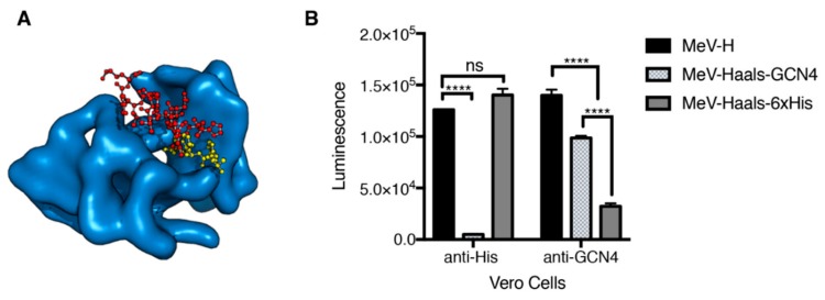
Effect of affinity binding in pseudoreceptor fusion levels. (A) Crystallographic structures for the tested peptide–pseudoreceptor systems. Crystallographic structures for the scFv binder to GCN4(7P-14P) peptide (PDB [Protein Data Bank] ID 1p4b) with the position of the His-peptide binding are shown (based on structural overlay with the scFv-His peptide structure, PDB ID 1kTR). The scFv binder is shown in a blue surface representation. GCN4(7P-14P) and His peptide binders are shown as red and yellow balls and spheres, respectively. (B) A quantitative fusion assay was performed with baby hamster kidney cells bearing the indicated H protein with MeV-F. Vero cells that were transfected with the corresponding pseudoreceptor were used as effector cells. Haals indicates an H protein blind for the natural receptors. Maximum values over a 10-h experiment are shown, with mean (SD) of a representative experiment performed in triplicate. **** indicates p <.0001. MeV-F indicates measles virus–F glycoprotein; MeV-H, measles virus–H glycoprotein; ns, no significance, determined through 1-way analysis of variance with Bonferroni’s multiple comparison test (Used with permission of Mayo Foundation for Medical Education and Research).

