Figure 4.
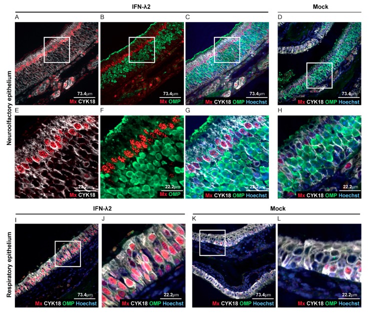
IFN-λ responsive cells in the olfactory epithelium. (A–L) Representative sections of the olfactory and respiratory nasal epitheliums labeled for Mx1 (red), olfactory marker protein (OMP) (neurons, green) and CYK18 (sustentacular cells, white). Where indicated, nuclei were stained with Hoechst dye (blue). Sections were from B6.A2G-Mx1-IFNAR1−/− female mice, seven days after electroinjection of an empty plasmid (n = 4) or a plasmid expressing IFN-λ2 (n = 4). (A–C,E–G) Representative sections of the olfactory epithelium of IFN-λ plasmid-treated mice revealing extensive Mx1 staining in sustentacular cells but not in neurons. (D,H) Little Mx1 expression in the olfactory epithelium of mock-treated mice. (E–H) Images zoomed-in from the upper panel (white square). (I–L) Extensive Mx1 expression in the respiratory epithelium of IFN-λ-treated mice (I–J) compared to mock-treated mice (K,L). (J,L) Images zoomed-in from the corresponding left panels (white square).

