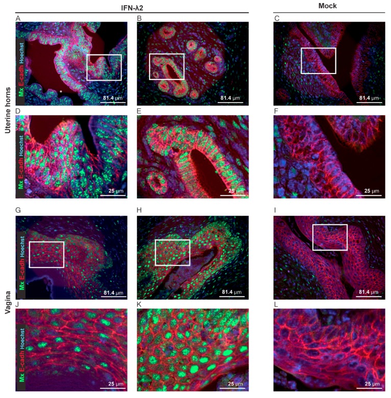
Figure 7.
IFN-λ-responsive cells in the genital tract. (A–L) Representative sections of the uterine horns and vaginas showing Mx1 expression (green) and E-cadherin+ epithelial cells (E-cadh, red) seven days after electroinjection of B6.A2G-Mx1-IFNAR1−/− female mice with an empty plasmid (Mock, n = 4) or a plasmid expressing IFN-λ2 (n = 4). (A–F) Representative sections of the uterine horns revealing extensive Mx1 expression in the E-cadherin positive cells of two IFN-λ-treated (A,B) compared to the mock-treated (C) mice. (D–F) Images zoomed-in from the upper panel (white rectangle). (G–L) Representative sections of the vaginal epithelium revealing partial (G–J) versus extensive (H–K) Mx1 expression in the E-cadherin positive cells of two IFN-λ-treated mice and little Mx1 expression in the E-cadherin positive cells of mock-treated mice (I–L). (J–L) Images zoomed-in from the upper panel (white rectangle).

