Figure 3.
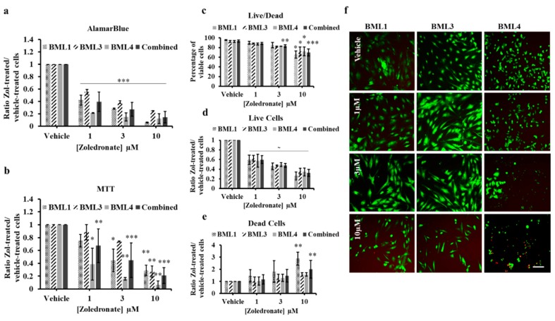
AlamarBlue (a) and MTT (b) assays of lung cancer-induced bone metastasis cells treated with vehicle (PBS1x) or Zol 1 µM, 3 µM and 10 µM for 7 days in 1% serum conditions. The histograms in (a) and (b) represent the ratio of drug-treated cells divided by vehicle-treated cells (PBS1x) in three independent experiments. (c) Percentage of viable cells [number of live cells/(number of live cells + number of dead cells) × 100] and (d) ratio of live cells or (e) ratio of dead cells in vehicle or Zol-treated conditions of Live/Dead assay carried out on bone metastasis cells. (f) representative photos of Live/Dead assay following vehicle or Zol treatment at different concentrations. Live cells are in green and dead cells are in red. Scale bar 250 µM. Significantly different from control * p < 0.05, ** p < 0.01, *** p < 0.001, ~ p values indicated in the RESULTS text.

