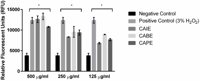
Figure 2.
Quantification of intracellular reactive oxygen species in the presence of three inhibitory compounds. Paenibacillus larvae were stained with 10 µM 2′,7′-dichlorodihydrofluorescein diacetate (H2DCFDA) before incubating the cells in the presence of minimum inhibitory concentrations of CAIE, CAPE, CABE and 3% H2O2 (positive control) for 18 h incubation at 37 °C. Fluorescence was quantified using a microplate reader at 495 nm/527 nm (Ex/Em). Data presented are mean RFU ± SD (n = 2–6). Asterisks represent a significant difference between control cells and cells exposed to either CAIE, CAPE, CABE and 3% H2O2 (p < 0.05).

