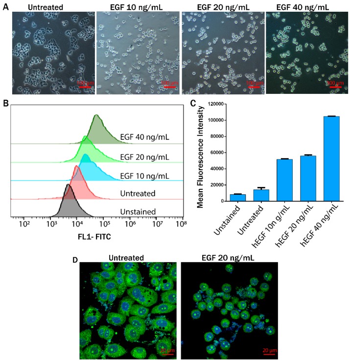
Figure 3.
Epithelial to mesenchymal transition (EMT) induction in MDA-MB-468 cells. (A) Changes in the morphology of cells treated with increasing concentrations of epidermal growth factor (EGF); (B,C) histograms from flow cytometry for vimentin expression (B), with corresponding mean fluorescence intensity shown in a bar plot (C); (D) alteration in the cytoplasmic and nuclear morphology studied by calcein-AM DAPI (4’,6-diamidino-2-phenylindole) staining.

