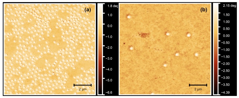
Figure 1.
Atomic force microscopy (AFM) phase images of the PNIPAM–co–AAc core microgels (a) and, as a typical example, the core-shell microgel sample PNN50/PMAM50 (b). In (b), the corona formed in the second precipitation polymerization step can be clearly identified. All measurements were performed in the dry state.

