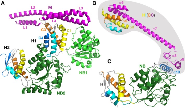
Figure 7.

Crystal structures of fragments of TtClpB chaperone and EcLon. (A) TtClpB chaperone (150–854; PDB ID 1QVR); (B) EcLon (124–245; PDB ID 3LJC); (C) EcLon (235–584; PDB ID 6N2I). The nucleotide‐binding domains are colored in light (NB1, TtClpB) and dark green (NB2, TtClpB and NB, EcLon). Corresponding helices in the H1 and H2 domains of TtClpB, as well as in HI(CC) and H domains of EcLon, are colored identically.
