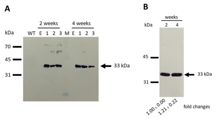
Figure 5.
Western blot analysis of empty vector and SynMAT expressing cells. (A) wild type, empty vector and SynMAT (2 and 4 weeks old). (B) SynMAT (2 and 4 weeks old). An equal amount of crude extract (20 µg) was loaded in all lanes. Blotting was conducted with a PVDF membrane. Antibody raised against 6x His-tag and HRP-conjugated anti-mouse IgG were used as primary and secondary antibodies, respectively. The membranes were developed using a using Horseradish Peroxidase Conjugate Substrate kit.

