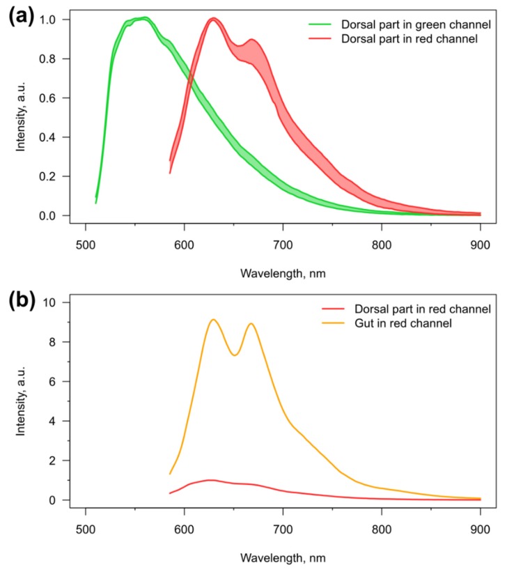
Figure 3.
Autofluorescence of E. verrucosus. (a) Variability (n = 5) in autofluorescence spectra of the dorsal parts of the first two pereon segments in green and red channels, and (b) representative comparison of autofluorescence intensity between the dorsal and central (containing gut) parts of third pereon segment of the same individual in red channel. Spectra on the upper panel are aligned at the regions of peak intensity.

