Figure 6.
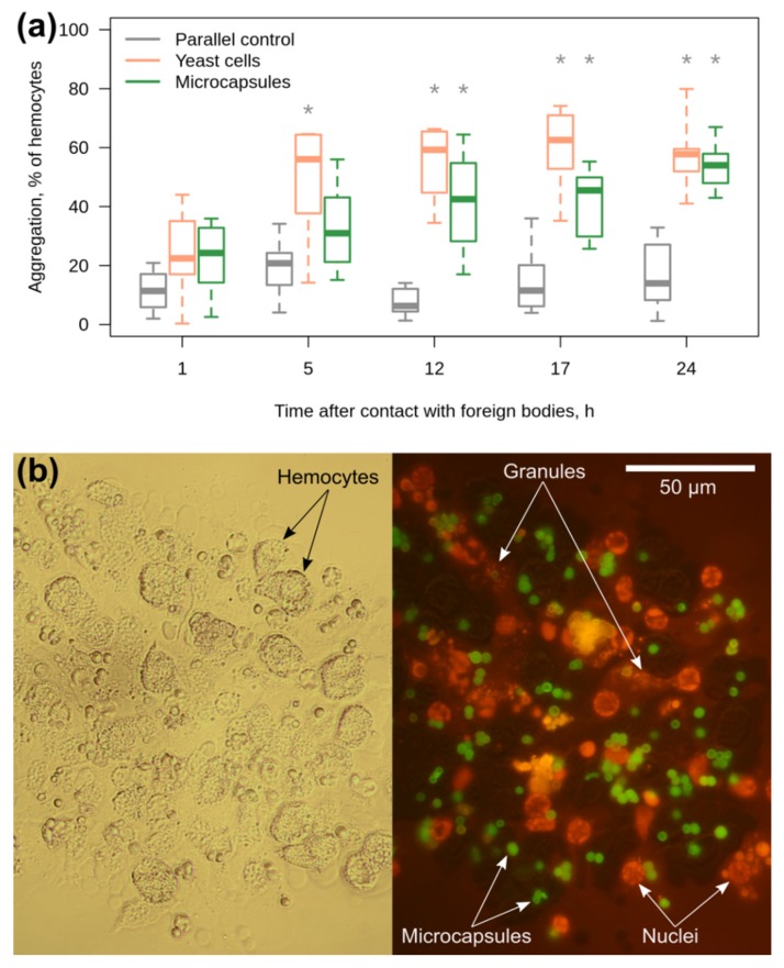
The reaction of primary culture of E. verrucosus hemocytes to yeast and microcapsules. (a) Monitoring of hemocyte aggregation after the introduction of yeast cells used as a positive control (1:1 of hemocytes) and microcapsules (5:1 of hemocytes) to the hemocyte media during 24 h (n = 8). (b) A representative example of a squashed aggregate of hemocytes with the microcapsules after the end of incubation in brightfield channel (left) and green fluorescent channel with the orange RuPhen3 staining (right). Microcapsules contained green FITC-albumin but partially obtained the yellow coloration due to contact with RuPhen3. * designates statistically significant difference from the parallel control group with p < 0.05.

