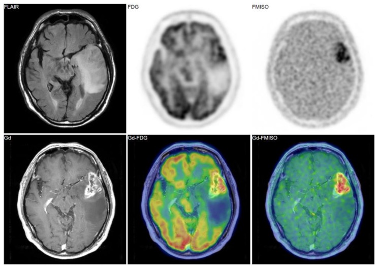
Figure 2.
A 69-year-old patient had a tumor in the left temporal lobe. Fluid-attenuated inversion recovery (FLAIR) image showed high intensity, indicating the tumor and the peritumoral edema. Gadolinium enhancement, 18F-fluorodeoxyglucose (FDG) uptake, and FMISO uptake were observed in the same area. The pathological diagnosis was glioblastoma (grade IV).

