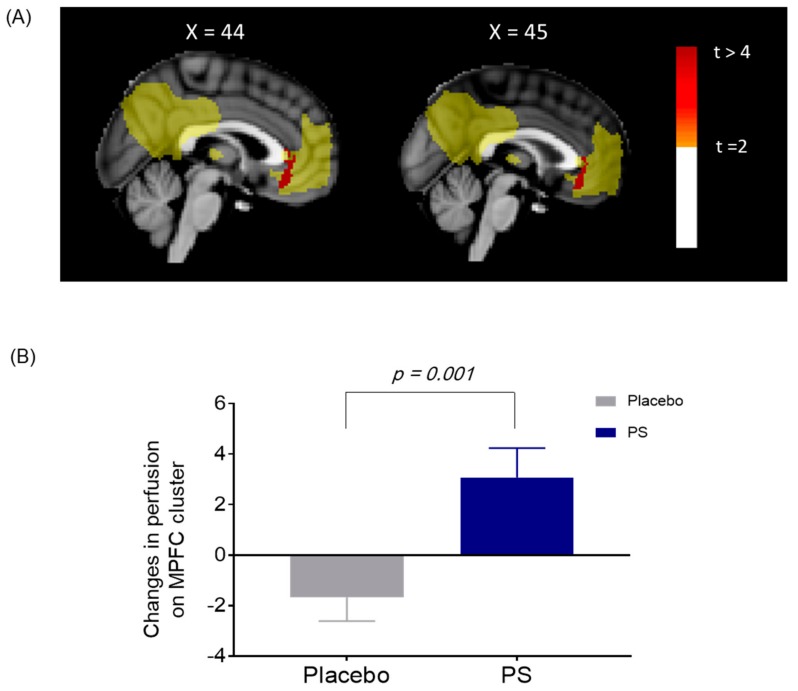
Figure 4.
(A) Changes in cerebral perfusion on the default mode network (DMN) between the PS and placebo group. Significant between-group differences in changes in cerebral perfusion within the medial prefrontal cortex (mPFC) of the DMN was shown after the four-week administration period (shown in red). (B) Increased cerebral perfusion on the mPFC in the PS group. The values were extracted from the mPFC for post-hoc analysis, which were significant clusters as shown in Figure 4 (A). Cerebral perfusion of the mPFC increased in the PS group, while decreased in the placebo after the four-week administration (z = −3.0, p for group-by-visit interaction = 0.001). mPFC, medial prefrontal cortex; PS, Polygonatum sibiricum

