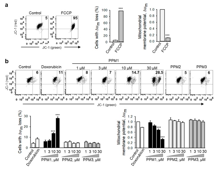
Figure 5.
PPM1 affects the mitochondrial integrity in breast cancer cells. (a) MDA-MB-231 cells treated with the respiratory uncoupler, FCCP (50 µM) for 6 h were used as positive control. (b) MDA-MB-231 cells were treated with doxorubicin (100 nM), different concentrations of PPM1, PPM2 or PPM3 (each at 30 µM) for 24 h. The mitochondrial membrane potential was analyzed flow cytometrically by using JC-1 dye. Representative dot plots are shown. Figures (upper right square) show number of cells with the loss of mitochondrial membrane potential (ΔΨm). ΔΨm was measured as red/green JC-1 fluorescence intensity ratio. Graphs demonstrate the percentages of MDA-MB-231 cells with depolarized mitochondria and the loss of ΔΨm in treated cells. Statistical analysis was performed by using the Newman-Keuls test. All data are mean ± SEM, n = 3, *** p < 0.001 vs. control.

