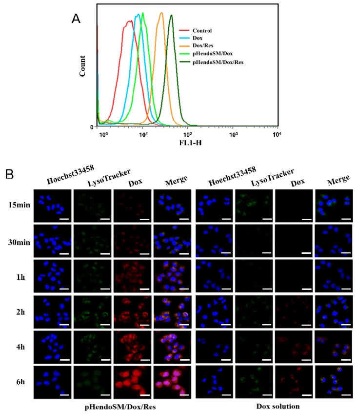
Figure 4.
Flow cytometry measurement of the intracellular uptake of Dox in the MCF-7/ADR cells treated with different formulations after 6 h incubation (A). The confocal laser scanning microscopy (CLSM) images of the MCF-7/ADR cells incubated with pH-endoSM/Dox/Res micelles and free Dox solution for 15 min, 30 min, 1 h, 2 h, 4 h, and 6 h at 37 °C (Dox concentration was kept at 2 μg/mL). Blue, green and red colors indicate Hoechst 33342, LysoTracker green and Dox, respectively. Scale bars represent 20 μm (B).

