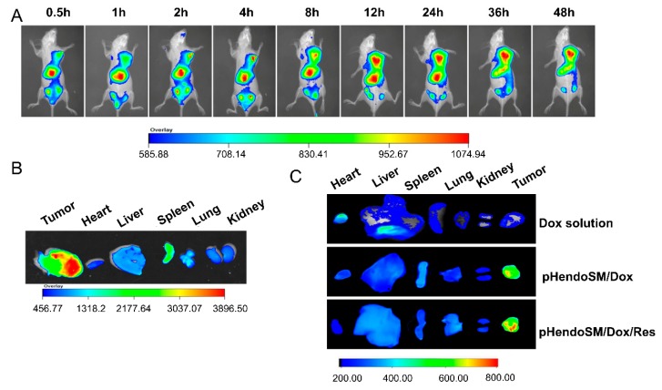
Figure 8.
In vivo imaging of MCF-7/ADR tumor-bearing nude mice after intravenous injection with DIR-loaded micelles (A), Ex vivo fluorescence images of tumors and organs collected at 48 h (B), In vivo distribution of Dox at 48 h after intravenous injected Dox solution, pH-endoSM/Dox, and pH-endoSM/Dox/Res (C) (n = 3).

