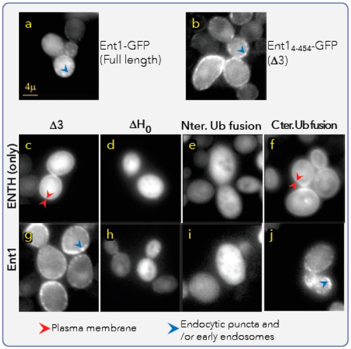
Figure 5.
Ent1 and Ub–Ent1 membrane binding assays in vivo. Fluorescent microscopy imaging of living yeast cells expressing GFP fusion of the indicated Ent1 derivatives are shown. (a) A full-length Ent1 with C-terminus in-frame fusion GFP, expressed from the native genomic promoter. (b) An Ent14-454 (Δ3) with C-terminus in-frame fusion GFP, expressed from a Gal promoter on a single copy plasmid. (c–j) The fluorescent readouts from indicated derivatives. ΔH0 is the construct that lacks the first 17 residues. In the Ub-fusion constructs, Ub was fused in-frame instead of K3 (i.e., to residue Q4). In the fusion-Ub constructs, Ub is fused in-frame at the C-terminus of the Ent1 derivative (between Ent1 and GFP). Red arrows mark probably endocytic or early endosome clusters of Ent1–GFP; yellow arrows mark the accumulation of Ent1-GFP fusions at the plasma membrane.

