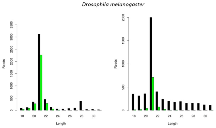
Figure 4.
Generation of virus-derived siRNAs in Drosophila melanogaster S2 cells. Length distribution of small RNAs mapping to the Drosophila A virus (DAV) (left) and to the Drosophila C virus (DCV) (right). Reads (y axis): number of reads. Length (x axis): length (nt) of the reads. Black: sense reads. Green: antisense reads. To avoid degradation products and general contaminants, reads shorter than 18 nt and longer than 31 nt were not included. Mapping was performed with Bowtie2, and graphs were obtained with the viRome package in R.

