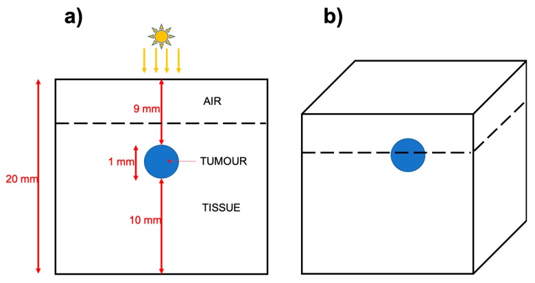
Figure 1.
Schematic showing the dimensions of the simplified skin cancer model. (a) A two-dimension (2D) slice through the simulated world, which is a cartesian grid illustrated in (b). The light source is 1 W and has a radius of 3 mm and is set to irradiate the top and centre of the model. In the simulations, we compare a control, to a tumour infused with gold nanorods, with the absorbance and scattering values (respectively μa and μa) shown in Table 1.

