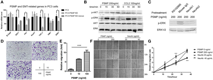
Figure 7.
Highly expressed PSMP in PC3 cells could promote EMT through p-ERK and p-AKT signaling pathway molecules. (A) After PSMP-deficient PC3 cell lines were established, mRNA was extracted from three different groups, including wild-type PC3 cells, PSMP-deficient PC3 cells, and PSMP-deficient PC3 cells cocultured with 50 ng/ml PSMP, and the expression of several EMT-related genes, including E-cadherin, ERβ1, FN1, Snail, TGF-β, VIM1, and ZEB1, was measured by qPCR. (B) After incubation with 100 ng/ml CCL2 or 200 ng/ml PSMP for 0, 15, 30, or 60 min, PC3 cells were collected, and phosphorylated AKT, total AKT, phosphorylated ERK1/2, and ERK1/2 levels in PC3 cells were detected by Western blotting. (C) PC3 cells were pretreated with 5 μg/ml RS102895, 500 ng/ml mIgG, 500 ng/ml neutralizing antibody 1, and 500 ng/ml neutralizing antibody 2 for 2 h, and then 200 ng/ml PSMP was added into the PC3 cell culture medium. After incubation for 30 min, phosphorylated ERK1/2 and ERK1/2 levels in PC3 cells were detected by Western blotting. (D,E) PBS or 10 or 100 ng/ml PSMP was added into the lower wells of Transwell plates, and PC3 cells were added to the upper wells with medium. After coculture for 48 h, PC3 cells that had migrated through the membrane were stained and counted. (F,G) PBS, 200 ng/ml PSMP, or 20 or 40 μg/ml neutralizing antibody was added into cell culture wells, and wound healing was observed. Then, cell migration was monitored during coculture for 36 h, images were captured, and the width of the wounds was measured at different time points.

