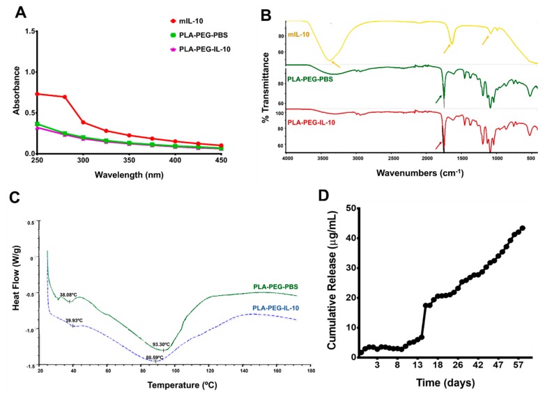
Figure 3.
Physiostructural characterization and release of nanoparticles. Nanoparticles and IL-10 were dissolved in DNAse-RNAse free water and analyzed by spectroscopy (A). FT-IR spectra were employed to show the various functional groups and differences in the absorption spectra for nanoparticles. The FT-IR spectrum was recorded with 64 scans/sample ranging from 4000 to 1000 cm−1 and a resolution of 4 cm−1 at ambient temperature (B). Thermal stability of IL-10 encapsulated within PLA-PEG nanoparticles was determined by placing nanoparticles in an aluminum pan, sealing it, and then heating it at the rate of 20 °C per min from 30 to 120 °C under nitrogen, before cooling it from 120 to 30 °C at the same rate, using a differential scanning calorimeter. Shown are peaks for PLA-PEG-PBS (green) and PLA-PEG-IL-10 (blue), respectively (C). The in vitro release of IL-10 from PLA-PEG nanoparticles was determined by placing nanoparticles in sterile PBS. At each time-point, supernatants were collected and the IL-10 content was measured spectrophotometrically. Shown is the average cumulative release of IL-10 over a period of 60 days, and the experiment was performed three times (D). DNA = deoxyribonucleic acid, IL-10 = Interleukin-10, FT-IR = Fourier Transform-Infrared, PBS = Phosphate-buffered saline, PLA-PEG = Poly (lactic acid)-Poly (ethylene glycol), RNA = ribonucleic acid, UV = Ultra-Violet.

