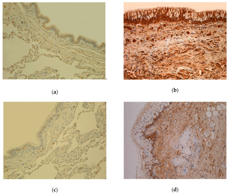
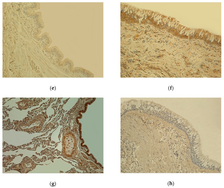
Figure 1.
(a) Weakly stained few to moderate number (+/++) IL-7-positive cells in bronchial epithelium and smooth muscle; few (+) IL-7 positive cells in connective tissue of 81-year-old female bronchial wall (Control). IL-7 IHC, X200. (b) Numerous to abundance (+++/++++) of IL-7-containing cells in bronchial epithelium and connective tissue of 61-year-old male lung tissue (chronic obstructive pulmonary disease, COPD). IL-7 IHC, X200. (c) Almost no positive (0) IL-1α-containing cells in epithelium and few (+) IL-1α positive cells in connective tissue of 81-year-old female bronchial wall (Control). IL-1α IHC, X200. (d) Few to moderate number (+/++) IL-1α immunoreactive cells in epithelium and connective tissue of 78-year-old male bronchial wall (COPD). IL-1α IHC, X200. (e) Weakly stained few (+) hBD-2-containing cells in bronchial epithelium and connective tissue of 81-year-old female bronchial wall (Control). hBD-2 IHC, X200. (f) Moderate number (++) of hBD-2 immunoreactive cells in bronchial epithelium and connective tissue of 78-year-old male bronchial wall; goblet cell hyperplasia (COPD). hBD-2 IHC, X200. (g) Numerous (+++) Hsp-70-containing cells in bronchial epithelium and cartilage; moderate number (++) of Hsp-70-positive cells in connective tissue of 81-year-old female bronchial wall (Control). Hsp-70 IHC, X200. (h) Almost no positive (0) Hsp-70-containing cells in bronchial epithelium and connective tissue of 60-year-old male bronchial wall (COPD). Hsp-70 IHC, X200.


