Figure 1.
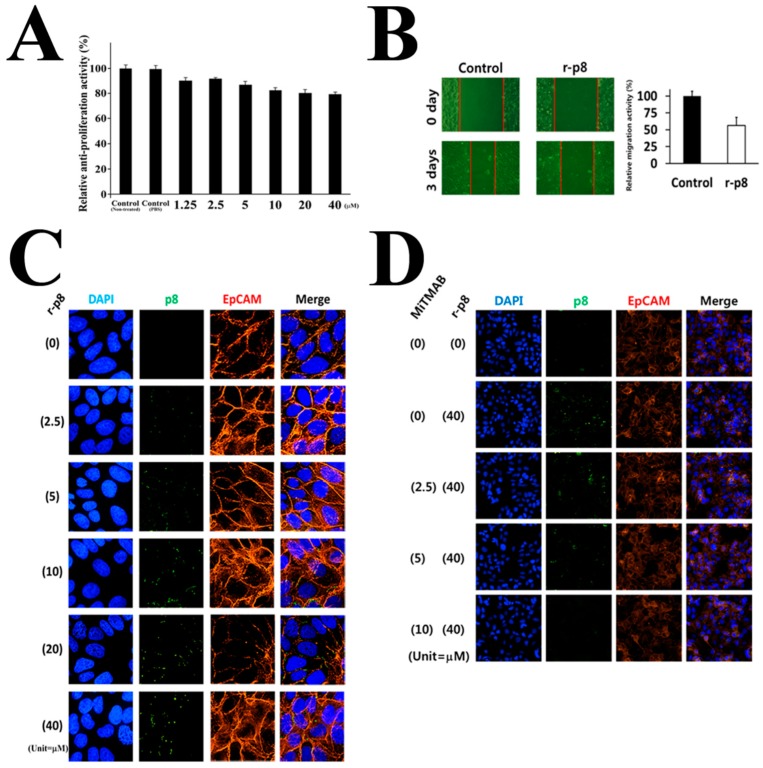
Characterization of p8 as an anti-cancer drug. Anti-cancer properties of exogenous r-p8 treatment. (A) R-p8 (0–40 μM) was incubated with DLD-1 cells (3 × 10³ cells/well) for 72 h, and anti-cancer efficacy was determined by MTT assay. (B) Anti-migration properties were examined in a wound healing assay. Wound healing was analyzed using Image J. (C) ImageXpress® Micro Confocal microscopy (60X) was used to determine the entry efficiency of r-p8. Entry of r-p8 into cells is concentration dependent. Cells were stained to detect r-p8 (Green), the cell membrane marker EpCAM (Red), or nuclei (DAPI: Blue). (D) ImageXpress® Micro Confocal microscopy (4X) was used to identify the route of entry used by r-p8. Cells were treated r-p8 (40 μM) with or without an endocytosis inhibitor (MiTMAB: 10 μM) and then stained to detect r-p8 (Green), the cell membrane marker EpCAM (Red), or nuclei (DAPI: Blue).

