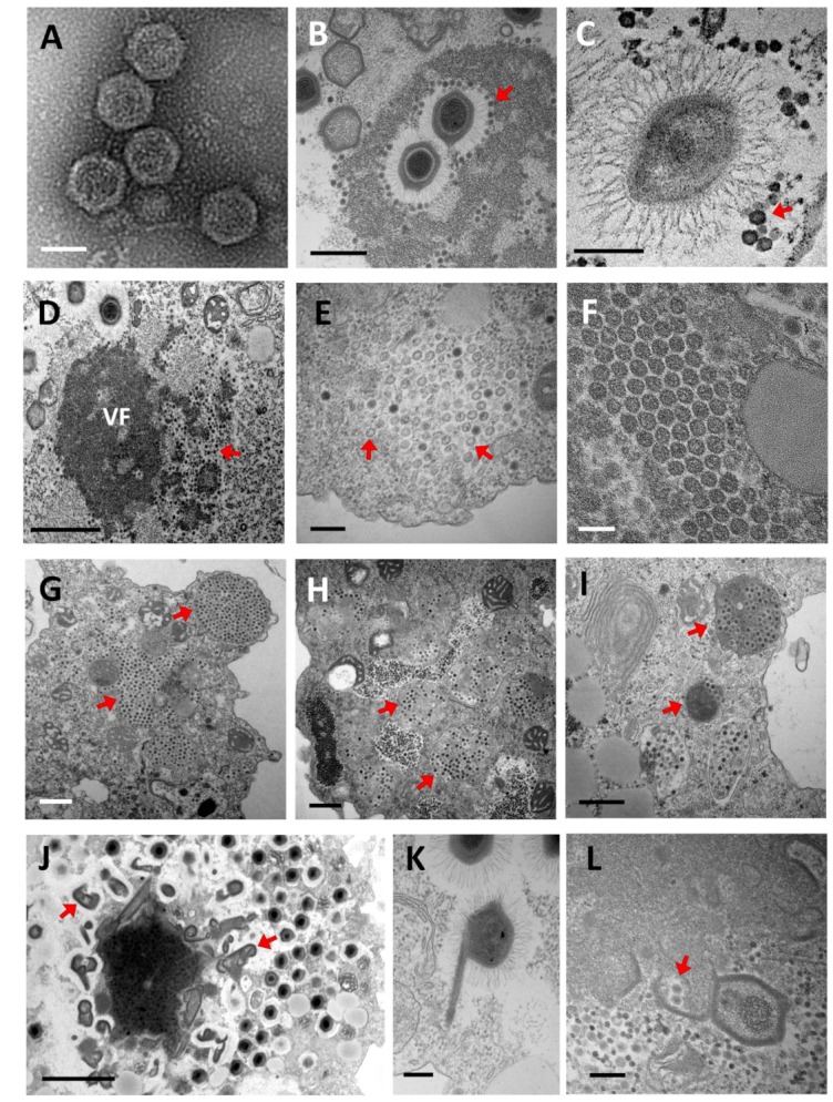
Figure 3.

Electronic microscopy observation of virophage particles and their replication cycle. (A) Negative staining electronic microscopy observation. (B–L) Transmission electronic microscopy images. (A) Morphology of purified Sputnik virophage particles (scale bar, 500 nm). (B,C) Sputnik and Guarani virions attached to the Mimivirus fibrils, respectively. (Scale bar, 500 nm and 200 nm). (D) Sputnik virophage invading the virus factory of acanthamoeba polyphaga Mimivirus (APMV) (scale bar, 1 µm). (E) Immature Sputnik virions observed in the cytoplasm of A. castellanii during co-infection with APMV (arrows) (scale bar, 200 nm). (F) Mature Sputnik virions (scale bar, 100 nm). (G–I) Virophages virions are commonly observed clustered inside typical cytoplasmic vesicles at the end of their replication cycles (arrows). (G) Sputnik progeny. (H) Zamilon progeny. (I) Guarani progeny (scale bars, 500 nm). (J,K) The genesis of abnormal Mimivirus particles has been observed during infection with virophages (arrows). (scale bars, 2 µm and 200 nm). (L) Encapsidation of virophage virions within the Mimivirus capsid (arrows) (scale bar, 200 nm). VF: Virus factory.
