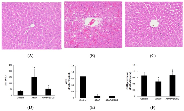
Figure 2.
Effects of EGCG supplementation (0.6%) on APAP-induced hepatotoxicity in rats. Histopathological examination of livers was shown in (A) the control group, (B) the APAP group, and (C) the APAP + EGCG group. H&E stain, 400×. Normal architecture of the liver was found in the control group (A). Multifocal necrosis was graded as slight (2) in (B) (APAP group) and minimal (1) in (C) (APAP + EGCG group). Plasma alanine aminotransferase (ALT) activity and hepatic reduced- glutathione (GSH) and GSH peroxidase activity were shown in (D–F), respectively. * Significantly different from control group, p < 0.05. # Significantly different from APAP group, p < 0.05.

