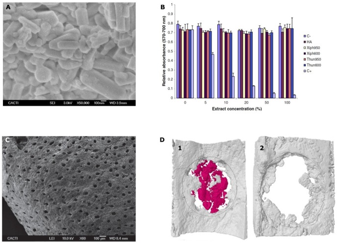
Figure 1.
(A) FSEM micrograph showing the appearance of the obtained powders from fish bones at 600 °C; (B) cell viability of osteoblasts (MC3T3-E1) after incubation with sword fish (Xiph) or tuna (Thun) samples treated at 600 or 950 °C together with commercial hydroxyapatite (HA) and control extracts for 24 h. C+: positive control. C−: negative control; (C) SEM micrographs of the shark teeth bioapatites morphology at 60×; (D) micro-CT reconstructions of an extracted bilateral parietal rat bone defect either treated with shark teeth bioapatites (1) or correspondent critical defect control (2) after 3 weeks of implantation. Marine shark teeth bioapatites are colored in red and new bone tissue in gray. Areas of interest are delimited by gray lines. Figure 1A,B reprinted from [76] with permission from Elsevier. Figure 1C,D reprinted from [78] with permission from John Wiley and Sons.

