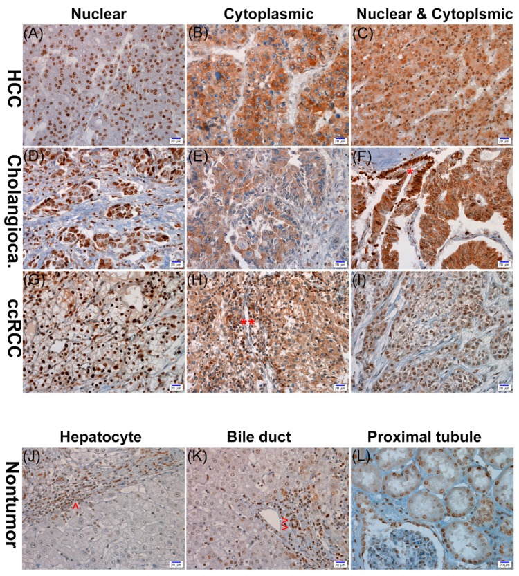
Figure 1.
Representative photographs of APEX1 immunohistochemical staining in hepatocellular carcinoma (HCC) (A–C), cholangiocarcinoma (Cholangioca) (D–F), and clear cell renal cell carcinoma (ccRCC) (G–I). The three cancers show nuclear, cytoplasmic, or nuclear and cytoplasmic expression of APEX1. The nontumor proliferative biliary epithelium (*) expresses nuclear staining, while the cholangiocarcinoma cells show both nuclear and cytoplasmic expression (F). Inflammatory cells (**) exhibit cytoplasmic staining (H). No APEX1 expression in nontumor hepatocytes in contrast to the high nuclear expression in inflammatory cells (^) (J). Positive nuclear expression in nontumor bile duct epithelial cells (^^) (K). Positive nuclear expression in proximal convoluted tubular epithelial cells (L) (scale bar = 20 μm).

