Figure 3.
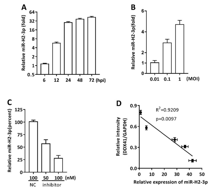
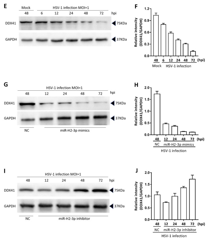
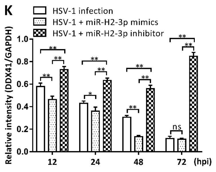
miR-H2-3p contributed to the downregulation of DDX41 during HSV-1 infection in THP-1 cells. (A–C) THP-1 cells were infected with HSV-1 at an MOI of 1 for indicated time point (A) or at an MOI of 0.01, 0.1, and 1 for 24 h (B), or transfected with miR-H2-3p inhibitor or negative control inhibitor at the indicated final concentration, and then infected with HSV-1 at an MOI of 1 for 24 h (C). The cells were harvested, and total RNA was extracted for determining the miR-H2-3p expression using qRT-PCR. The values were normalized to U6 sno RNA and standardized to 1 in 6 hpi (A) or an MOI of 0.01 (B) or NC treatment (C). (D) Analysis of correlation between miR-H2-3p expression (A) and DDX41 expression (E) during HSV-1 infection with an MOI of 1. R, Reliability; p, Pearson correlation coefficient. (E and F) THP-1 cells were infected with HSV-1 at an MOI of 1 or mock (Mock) for indicated time periods. The level of DDX41 protein was determined by immunoblotting (E) and the relative density of DDX41 protein using ImageJ software (F). The values were normalized to GAPDH, using GAPDH as a loading control. (G–J) THP-1 cells were transfected with miR-H2-3p mimics or negative control mimics at the final concentration of 100 nM or miR-H2-3p inhibitor or negative control inhibitor at the final concentration of 100 nM, and then infected with HSV-1 at an MOI of 1 for 24 h. The level of DDX41 protein was determined by immunoblotting (G,I) and relative density of DDX41 protein using ImageJ software (H,J). The values were normalized to GAPDH, using GAPDH as a loading control. (K) Summary for panel of F, H, and J. Data are the means ± SD (n = 3) from one representative experiment. Similar results were obtained from three independent experiments. * P < 0.05; ** P < 0.01.


