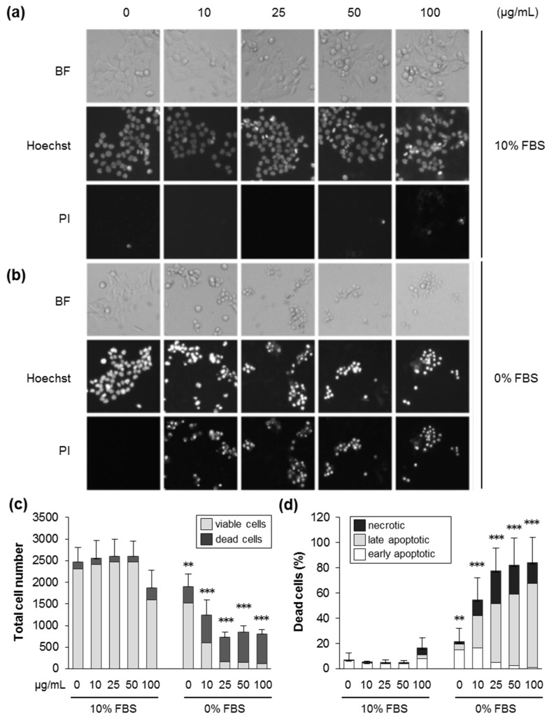
Figure 2.
Silica NPs induce apoptotic and necrotic cell death in the absence of serum. HCT116 wt cells were incubated with SiO2—12 nm NPs at the indicated concentration in the presence (10% FBS) or absence (0% FBS) of serum. After 24 h, the cells were stained with Hoechst and propidium iodide (PI), and images were acquired by automated microscopy and analyzed by the scan^R software to deduce cell numbers and the different modes of cell death. (a,b) Representative images of cells treated as indicated in the brightfield, the Hoechst, and the PI channels. (c) The total cell number divided into living and dead cells after treatment, as indicated. (d) The percentage of dead cells relative to the total cell number divided into the different classes of cell death, as indicated. Data are represented as mean values of three independent experiments carried out with four replicates (n = 12). The error bars are SD values related to the total cell number (c) or the percentage of dead cells (d). ** p < 0.01, *** p < 0.001 indicate significant differences in the response of cells treated with particles at corresponding concentrations in the absence (0% FBS) or the presence of serum (10% FBS).

