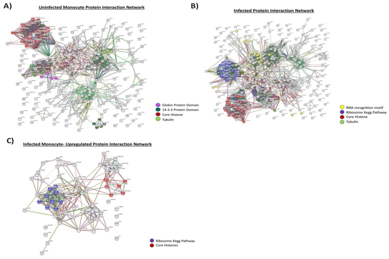
Figure 3.
Protein comparison of uninfected and infected monocyte EVs. 5-day old cell supernatants from U937 (uninfected) and U1 cells (HIV-1-infected) were harvested, treated with ExoMAX overnight, and centrifuged. The resulting pellet was run on an iodixanol density gradient, and the 10.8 fraction (exosome fraction) [36] was treated with NT80/82 overnight. The resulting pellet was treated was then prepared for mass spectrometry, and the resulting peptides were identified using Proteome Discoverer software. The predicted protein–protein interactions generated following multiple proteins input into the STRING database. A high confidence cutoff of ≥0.70–0.90 was implemented in this work. Network nodes represent proteins. Edges represent protein-protein associations and color shows association types. (A) Protein interaction network of proteins derived from uninfected monocyte exosomes (U937). (B) Protein interaction network of proteins derived from infected monocyte exosomes (U1). (C) Protein interaction network of proteins which are upregulated in infected monocyte exosome.

