Figure 4.
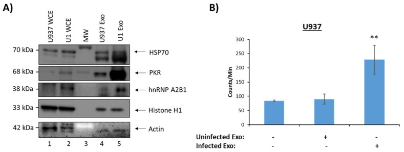
Infected monocyte EVs functional effects on recipient cells. 5-day old cell supernatants from U937 (uninfected) and U1 cells (HIV-1-infected) were harvested, treated with ExoMAX overnight, and centrifuged. The resulting pellet was run on an iodixanol density gradient, and the 10.8 fraction (exosome fraction) [36] was treated with NT80/82 overnight. (A) Samples were Western blotted for HSP70 (control), PKR, hnRNPA2/B1, histone H1, and actin (control). (B) Isolated EVs were added to recipient uninfected U937 cells, which had been synced at G0 phase, at a ratio of 1 cell:500 EVs and incubated for 44 h. [3H] thymidine was incorporated into the cells during a 4 h incubation. Cells were then washed and counted in a beta-counter to determine DNA synthesis. Student’s t-test compared untreated cells with cells treated with exosomes. ** p < 0.01, Error bars, S.D.

