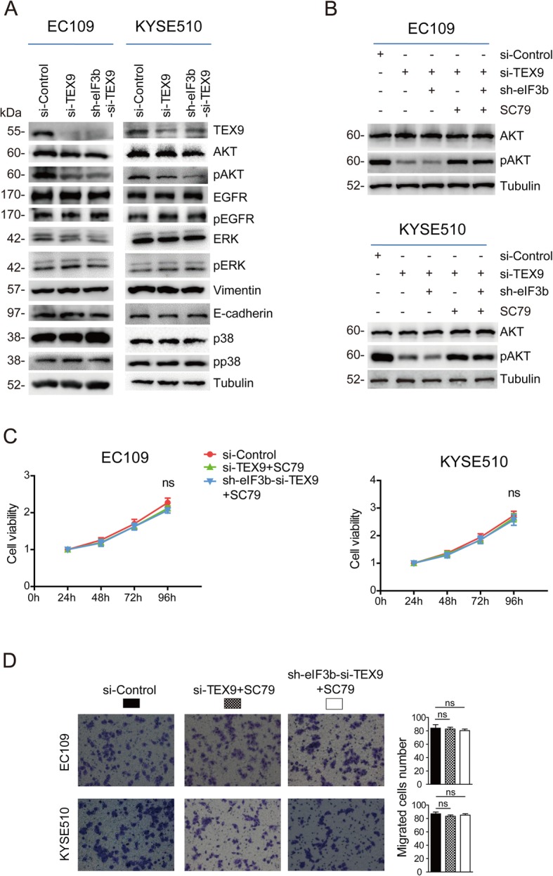
Fig. 4.

TEX9 and eIF3b promote ESCC progression through the activation of AKT signaling pathway. a Western blot assay was performed to analyze the expression difference of the related protein after depletion of eIF3b and TEX9. Tubulin was used as an internal reference. b Western blot assay was performed to analyze the AKT and pAKT expression after the treatment of SC79. The cells were starved for 30 min and treated with SC79 (10 μg/mL) for 1 h prior to the extraction of protein. c CCK-8 assay was performed to detect the reversed effect on proliferation after the treatment of SC79 for 24 h. d Transwell assay was performed to detect the reversed effect on migration after the treatment of SC79 for 24 h. (ns: no significance, *p < 0.05, **p < 0.01, ***p < 0.001)
