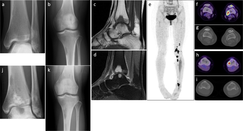
Fig. 1.
Imaging findings at presentation. a and b Plain X-ray findings. Multifocal ill-circumscribed lytic lesions were seen in the left femur, patella, tibia, and talus. c and d Sagittal magnetic resonance imaging findings of the left ankle. The multifocal lesions showed c homogeneous low intensity on T1-weighted images and d homogeneous enhancement after gadolinium administration. e-i Fluorodeoxyglucose (FDG)-positron emission tomography/computed tomography showed multifocal FDG-avid lesions in the bones of the left lower extremity with a maximum standardized uptake value of 15.95. No lesions were found in the soft tissue. j and k Rapid progression after curettage and artificial bone grafting of the lesion at the distal end of the left tibia was observed. Each ill-circumscribed lytic lesion in the left extremity increased in size over 1 month

