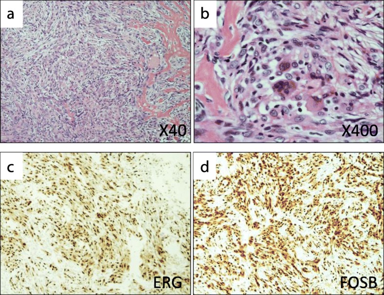
Fig. 2.

Pathological findings. a and b Hematoxylin–eosin staining showed proliferation of spindle and epithelioid cells with eosinophilic cytoplasm, which was compatible with the diagnosis of pseudomyogenic hemangioendothelioma. c and d Immunohistochemical studies showed that the tumor cells were positive for ERG and FOSB
