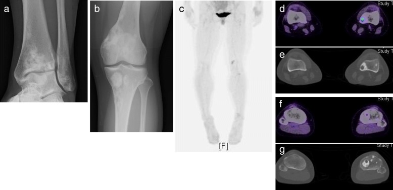
Fig. 3.
Imaging findings 4 years following denosumab treatment. a and b Plain radiographs showed shrinkage of the lesions with striking marginal sclerosis. c-g Fluorodeoxyglucose (FDG)-positron emission tomography/computed tomography showed that each lesion had become less FDG-avid with a maximum standardized uptake value of 2.6 and was accompanied by remarkable sclerotic bone

