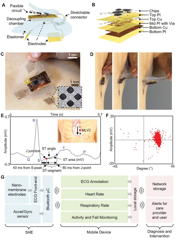
Figure 1.

Overview of the SHE and data management. A) Schematic illustration showing the main structural components and their assembly for SHE. B) Blow‐up rendering image detailing the layer information of the flexible circuit. C) Photo of an SHE showing the soft and adhesive property for direct lamination with the skin without the use of adhesives. D) Side view of delamination sequence of the ultrathin device with no negative effects to the skin. E) Detail inspection of ECG acquired by the SHE in (C) and identification of PQRST waves, J‐point, and ST‐segment angle. Inset diagram describes the position of SHE (red dot) and polarity of the two measurement electrodes. F) Scatter plot showing the angle and amplitude distribution of ST‐segments of the ECG data in (E). G) Flow‐chart illustration of a real‐time, smart, and ambulatory health monitoring, enabled by the SHE.
