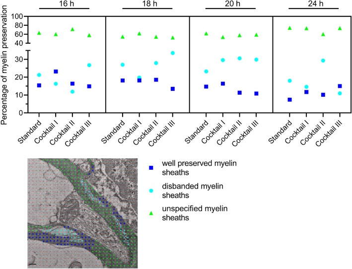
Fig. 3.
Quantification of the quality of the preserved myelin sheaths with different substitution cocktails compared to standard embedding. Analysis of the quality of the myelin sheath preservation, well-preserved myelin sheaths in blue squares, disbanded myelin sheaths are cyan dots, and unspecified myelin sheaths are shown in green triangles. Representative EM image with a red point grid and the color-marked distinction of the myelin sheaths, scale bar 0.2 μm

