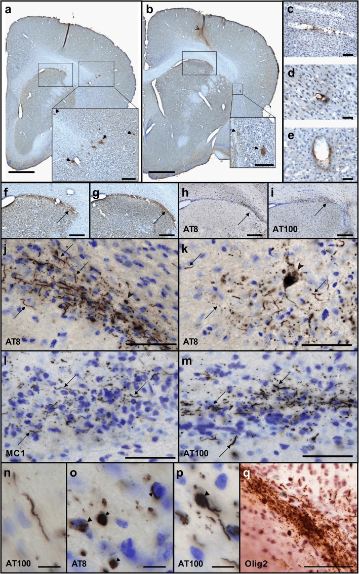
Fig. 5.
Amyloid depositions and tau inclusions in Alzheimer-inoculated mouse lemurs. a, b β-amyloid plaques (insets, arrows) revealed in two brain sections, immunostained for Aβ (4G8), separated from 1 mm in an animal from the Alzheimer-inoculated group. The inoculation site is shown with an open arrow (b). β-amyloid could also be detected in blood vessels (c-e) as well as in the parenchyma (f-g) close to the corpus callosum (arrows, images in f-g correspond to frames in a-b). Immunostaining for tau lesions using AT8 (h, j, k, o), MC1 (l) and AT100 (i, m-n, p). h-i frames display the same regions as the ones shown in (f-g): tau lesions (arrows) were detected in the same regions as Aβ. j-n are magnified images showing tau in neuropil threads (arrows). Intracellular tau positive structures were also detected in the form of globular positive cells (arrow), horseshoe intracellular accumulation (dotted arrow) and punctiform intracellular accumulation (arrowhead) (o). Rare somatodentridic inclusions (arrowhead k, p) as well as immunoreactive neurites with varicosities or "strings of beads" labeling (k: dotted arrow) were also detected. Tau stainings were counterstained with cresyl violet to identify neurons in (h-p). q displays AT8 sections double-stained for oligodendrocytes. Tau-positive lesions were not colocalized with oligodendrocytes (AT8 (brown) and Olig2 (red)). Scale bars: 1 mm (a-b), 200 μm (insets in a-b, e-i), 50 μm (c-e, j-m, q), 10μm (n-p)

