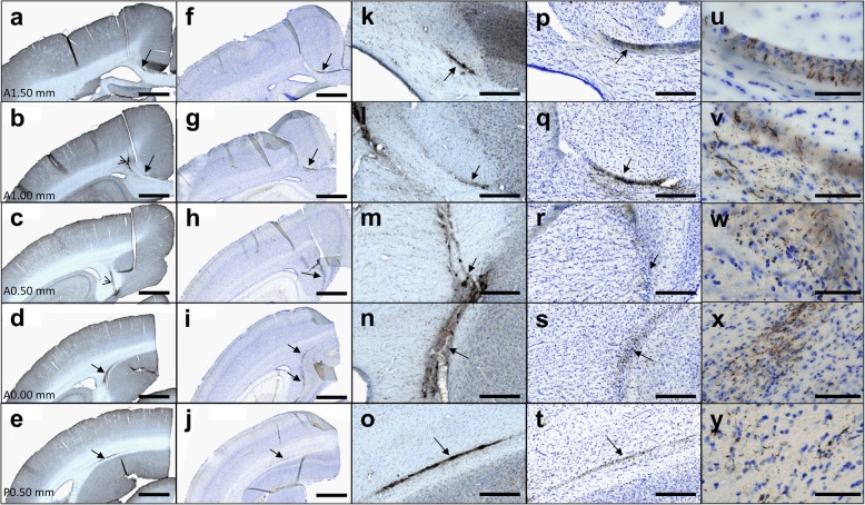
Fig. 6.
Diffusion of β-amyloid deposits and tau inclusions in Alzheimer-inoculated mouse lemurs. Immunostaining of Aβ (4G8, a-e, k-o (magnified views)) and tau (AT8, f-j, p-y (magnified views)) in 5 successive brain sections. u-y displays magnification of the tau-positive lesions from (f-j) or (p-t). β-amyloid and tau deposits (a-t) were seen exactly at the same locations (arrows). They spread from the inoculation site (open arrow in b, c) to regions localized one millimeter ahead and behind the inoculation site (A1.50 mm to P0.50 mm correspond to spatial references in the Bons atlas [31]). Scale bars: 1 mm (a-j), 200 μm (k-t), 50 μm (u-y)

