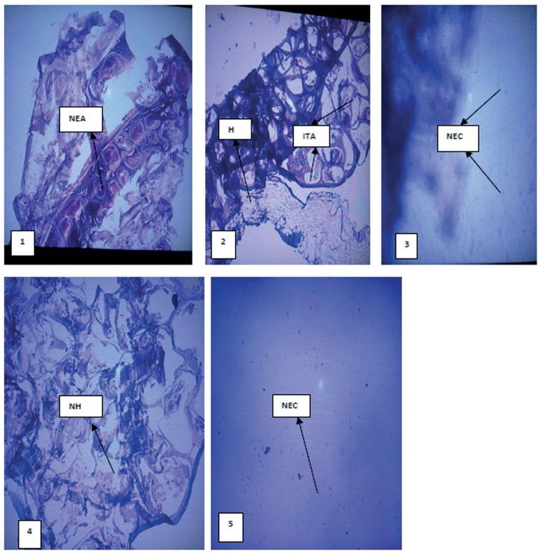
Figure 7.
Photomicrograph of the epididymis: 1 (H2O), 2 (Pb alone), 3(Pb+750mg/kg CA), 4 (Pb+1500mg/kg CA) and 5 (Pb+2250mg/kg CA). All panels were stained with hematoxylin & eosin, magnification x100. NEC (normal epididymal cell), NEA (normal epididymal architecture), ITA (inflamed tunica albuginea), H (hydrocele)

