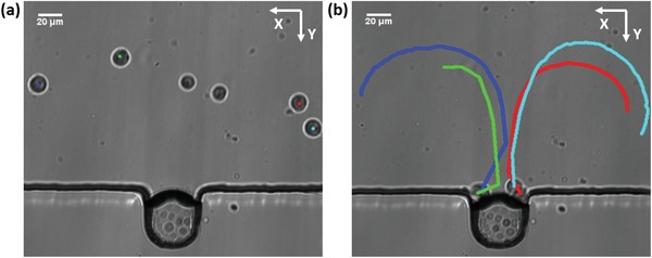
Figure 3.

Cells are trapped by the microbubble oscillations. a) Prior to the application of ultrasound, the cells are distributed randomly in the channel. The solution of harvested MDA‐MB‐231 cells is injected to the microchannel. b) Moving trajectory of the cells within the microstreaming. With the presence of ultrasound, cells are attracted to the proximity of the bubble membrane and trapped there.
