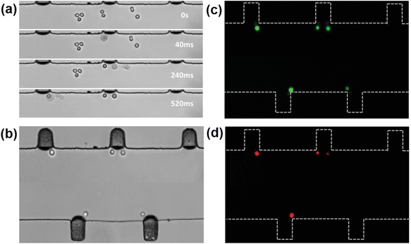
Figure 5.

a) With the presence of ultrasound, all cells in the microchannel are attracted toward the oscillating microbubbles in 520 ms. b) Bright‐field image of a single cell trapped at an oscillating microbubble surface. c,d) Fluorescence images of the membrane permeability of 88.89 ± 1.53% cells are enhanced by their corresponding oscillating microbubble and almost all the cells remain viable (n = 5).
