Figure 5.
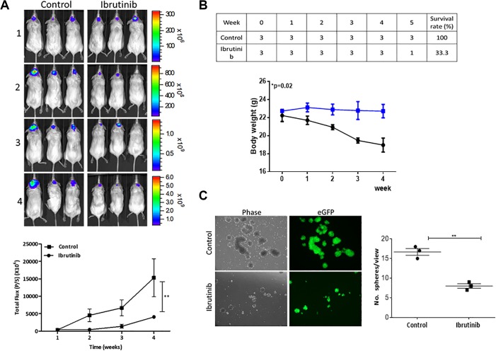
Preclinical evaluation of anti-glioma stem cell (GSC) function of ibrutinib using noninvasive bioluminescence imaging. A, Representative bioluminescence images of mice bearing DBTRG-05MG-CD133-LG neurospheres with and without the treatment of ibrutinib (over the course of 5-week experiment [Ib; intraperitoneal, IP, injection; 6 mg/kg; 5 times/week]) over time. N = 3 per group. Lower panel, Average body weight over time curve shows the treatment of Ib did not significantly affect the body weight of the animals. Lower panel represents the semiquantitative analysis of tumor burden over time. Y-axis, total photo flux; X-axis, time (treatment time). B, Survival rate and body weight monitoring over time. C, Comparative neurosphere-forming ability assay. Tumor samples were harvested from both control and Ib-treated mice. Ib-treated cells formed a significantly lower number of neurospheres, corresponding with a lower percentage of GFP+ cell population. *P ≤ .05; **P ≤ .01. Ib indicates ibrutinib; GFP, green fluorescent protein.

