FIGURE 1:
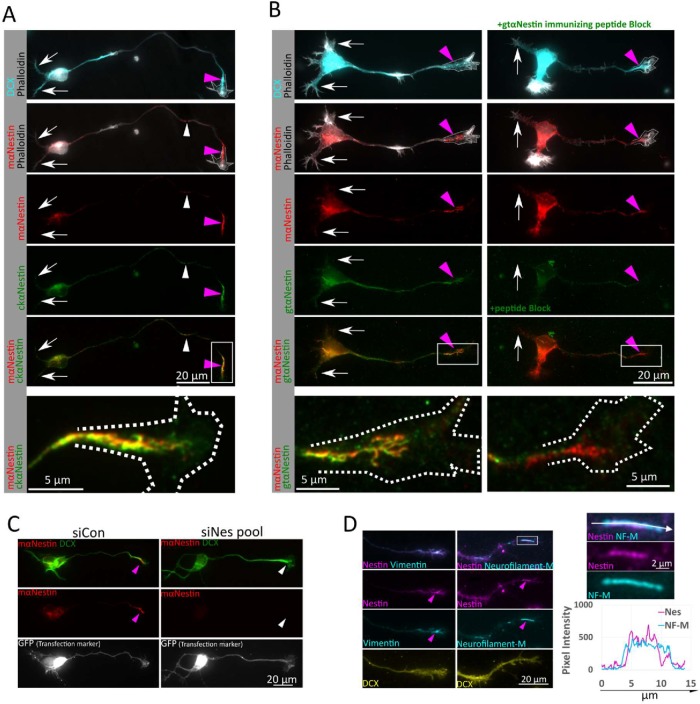
Nestin protein expression persists in immature primary cortical neurons in culture. (A). Nestin expression in cultures derived from E16 mouse cortex grown for 1 d (1DIV). Doublecortin (DCX) immunostaining and a stage 3 morphology were used to identify neurons. Nestin is enriched near the axonal growth cone (pink arrowheads), but largely absent from growth cones of minor dendrites (white arrows) using both mouse and chicken anti-nestin antibodies. White arrowheads indicate short nestin intermediate filament segments found along the axon shaft. Bottom panel is enlarged inset of growth cone, rotated, from within the white box. Growth cone outline drown from the phalloidin stain shown in the panels with phalloidin and in the enlargement. (B) Validation of nestin antibody staining on stage 3 neurons with mouse and goat anti-nestin antibodies with immunizing peptide antibody blocking. Both antibodies again show a distal axon enrichment of nestin (pink arrowheads), while the dendrites again are not labeled (white arrows). Preincubation of the goat anti-nestin antibody with the immunizing peptide abolishes all staining, but staining in the distal axons is still detectable with the mouse anti-nestin antibody, which was raised against a different epitope. Shown are E16 mouse cortical neurons 1DIV. Bottom panel is enlarged inset of growth cone from within the white box. Growth cone outline drawn from the phalloidin stain shown in the panels with phalloidin and in the enlargement. (C) Nestin siRNA results in loss of immunostaining, validating that the axonal growth cone staining is due to nestin protein. GFP was cotransfected as a transfection indicator. After 36 h in culture, cells were fixed and immunostained for nestin and DCX. The number of GFP positive cells positive for nestin in the axon as a fraction of the number of cells counted was quantified. (D) Nestin is colocalized with other intermediate filaments in the same assembly group (vimentin and Neurofilament-Medium NF-M) in the distal axons (pink arrowheads) of E16 mouse cortical neurons 1DIV. NF-M expression was low in immature cortical neurons cultured for only 1DIV. High magnification of the boxed region and line scans reveal that nestin and NF-M appear to decorate the same filament structure in partially colocalizing subdomains, as expected for nestin-containing intermediate filaments. Pink arrowheads indicate nestin-positive axonal intermediate filament heteropolymers.

