FIGURE 2:
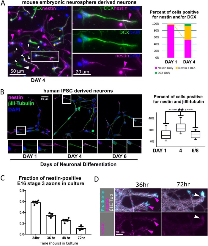
Nestin-expressing neurons are observed in multiple rodent and human culture models. (A) Mouse neurosphere NPC cultures: nestin expression was assessed in differentiating dissociated neural progenitor cells (mouse NPC) after 4 d of neuronal differentiation. DCX-positive neurons frequently stain positive for nestin (yellow asterisks), especially in axon tips (pink arrowheads). An example of an axon positive for DCX but not nestin is marked with a series of white arrowheads along the length of the long and thin axon, and a large white arrowhead indicating the growth cone. Nestin–single positive cells are marked with pink asterisks. One example of a double-positive cell is shown in the middle panels to highlight its neuronal morphology and tip-enriched nestin staining (pink arrowhead). The proportion of cells positive for only nestin (red bar) or only DCX (green bar) or double-positive for DCX and nestin (yellow bar) was quantified for NPCs cultured under differentiation conditions from 1 or 4 d. Yellow dotted lines highlight the expanding double positive population (yellow bar). Three hundred twenty-three cells were counted at 1 DIV, and 219 cells at 4 DIV. (B) Human IPSC–derived neuronal cultures: nestin is expressed in human (IPSC) differentiating neurons derived from NPCs in dissociated cultures under differentiating conditions from 1, 4, and 6 d. The distribution of nestin is similar to that in the mouse NPC–derived neurons described above. Percent colocalization with the neuronal-specific βIII tubulin was quantified. A close-up of a nestin-positive axon tip in the boxed region is shown in the panels below the respective images. n = 14 (day 1), 10 (day 4), and 14 (day 6–8). (Statistics: Mann–Whitney t test). (C) Mouse primary neuron cortical neuron cultures: percentage of nestin-positive neurons decreases rapidly with time in culture (30–60 stage 3 neurons were counted per time point for 3–5 experiments, as shown as the n). Cortical neuron cultures were prepared from E16 mice embryos and culture times are indicated. (D) Mouse primary explants: axonal nestin expression is progressively lost between 36 and 72 h in culture. Significant axonal nestin immunostaining is no longer detected by 72 h. E16 mouse cortex was explanted by incomplete dissociation and cultured. Axon fascicles emerging from the explant are shown. Arrowheads point to axon tips.

