FIGURE 3:
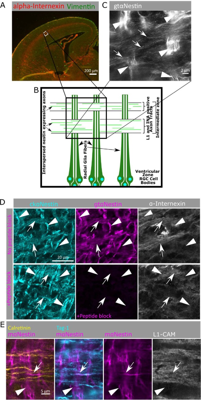
Nestin is expressed in subpopulations of cortical axons in developing cortex. (A) A low magnification of a coronal cortical section (E16 mouse) is shown for orientation (top) and showing α-internexin localization to the axon-rich intermediate zone. The white box (lateral lower intermediate zone) is the region imaged in the following panels, and is diagramed below (B), illustrating the vertically oriented radial glia and horizontal IZ axon bundles. (C) Superresolution microscopy (STED) of nestin immunostaining reveals bright staining of radial glial fibers (arrowheads) as well as fainter staining of axons (arrows) in mouse E16 cortex. Many axons in the axon fascicle do not express nestin, so only a subset of axons in the intermediate zone express nestin at this time point. Arrowheads indicate radial glia and arrows indicate nestin-positive axons. (D) Nestin staining of the lower intermediate zone of E16 mouse cortex using chicken anti-nestin (cyan) and goat anti-nestin (magenta) antibodies. Axon tracts are visualized with α-internexin antibody (white). Nestin staining is found in radial glia fibers (arrowheads) as well as in α-internexin-positive axon tracts (arrows). The goat anti-nestin antibody was preincubated with immunizing peptide on sequential cryosections in the lower panels. All staining with the goat anti-nestin antibody was blocked by peptide preincubation, including the axon tract staining, demonstrating that the axon staining was specific and not background staining. All images correspond to higher magnification of the lower intermediate zone of the lateral E16 mouse cortex (boxed regions in A). Radial glia are oriented vertically (arrowheads) and axon tracts are oriented horizontally (arrows). (E) Identification of nestin-positive axons in mouse cortex. The L1-CAM-positive axons (white) contain mixed populations of both cortical and thalamic projections in this brain region. Nestin (magenta) is specifically expressed in axons originating from the cortex (Tag1-positive, cyan), but not in axons with thalamic origin (calretinin-positive, yellow). Arrowheads indicate radial glia, and arrows indicate nestin-positive and Tag1-positive axons.

