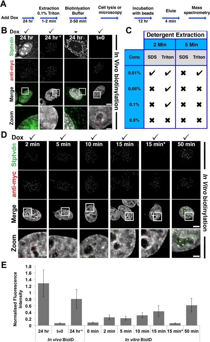
FIGURE 2:
Rapid in vitro biotinylation (ivBioID) of CENP-A. (A) Flowchart of ivBioID method. (B) Immunofluorescence of in vivo prepared BirA*–CENP-A cells. In panels B and E, 24 h+ represents an in vivo BioID sample extracted with 0.1% Triton X-100 before fixation. Panels t = 0 represent a sample with no biotin incubation. (C) Summary of results of cells prepared using the protocol described in A, under a variety of detergent extraction conditions. (D) Immunofluorescence of ivBioID–CENP-A prepared cells. Cells were permeabilized with 0.1% Triton X-100 extraction for 2 min. Several biotinylation buffer incubation time points were tested. Cells were fixed and processed for IF as standard and labeled with streptavidin 488 (green) or anti-myc (red). In panels D and E, 15 min* represents cells incubated with biotin buffer lacking ATP. Bar = 5 μm; Zoom bar = 1 μm. (E) Bar graph showing quantification of centromeric immunofluorescence from samples prepared as for B and D. Bars show the mean fluorescence of streptavidin 488 normalized to myc fluorescence. Ncell = 20. Ncentromere = 200.

