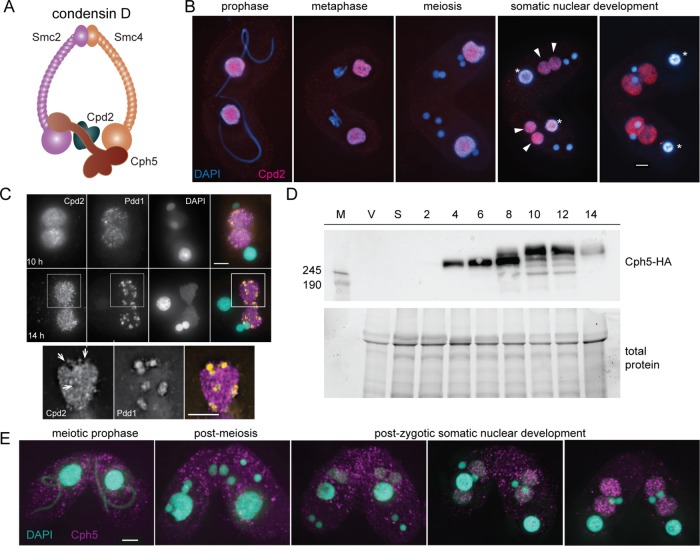
FIGURE 2:
Cpd2 and Cph5 localize to SN. (A) A developmental form of condensin (condensin D) is probably composed of Smc2, Smc4, Cpd2, and Cph5. (B) Cpd2-HA immunostaining (red) shows that Cpd2 localizes to paternal SNs during the early stages of mating. After fertilization and postzygotic divisions, Cpd2 appears in the differentiating SNs as they begin to swell (arrowheads), and remains in the developing nuclei until the late stages of mating, whereas it is lost from parental SNs (asterisks). (C) Costaining of Cpd2-HA and Pdd1 (orange) in developing SNs shows that Cpd2 is excluded from elimination bodies. Projected images of 3D stacks at 10 and 14 h. Enlarged images of a selected slice from the cell shows regions that lack Cpd2 staining (arrows) contain a Pdd1 signal in the merged image. (D) Western blots of whole cell protein extracts show that Cph5-HA is expressed as early as 4 h after initiation of mating, which corresponds to meiotic prophase. However, at later time points corresponding to SN development (8–14 h), additional Cph5-HA bands suggest that posttranslational modifications (probably phosphorylation) have occurred. M, molecular weight marker; V, vegetative cells; S, starved cells; 2–14, cells at the given number of hours after initiation of mating. (E) Immunostaining of Cph5-HA expressing cells fixed at different stages of mating shows that Cph5 specifically localizes to SN at late stages of development. Scale bars, 5 μm.

